Breast Cancer Screening Method

There will introduce breast cancer screening methods through using molybdenum sputtering target.
1. Imaging principle of mammography:
Use soft X-ray to shooting breast tissue, conducted by photographic film, after developing and fixing and other imaging procedures to form an image.
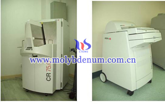
2. Major equipment of mammography X-ray machine: X-ray tube, breast compression device, grids, console
1) X-ray tube: it is a determining factor to obtain a high contrast image of the breast. General X-ray machine, anode target surface is tungsten, produces 0.008---0.031nm wavelength, wave length, penetration great, and it is hard rays. On the other hand, the wavelength of molybdenum produced 0.063---0.071nm wave length, long wavelength, penetration is weak, and it is soft rays. The wavelength produced by rhodium target is between the two, the penetrating is strong than molybdenum and display effect of dense glandular is better than molybdenum. The wavelength usually is 0.008---0.05nm.
2) the role of breast compression device:
a. Appropriate compression can reduce scattered rays for detection’s object contract.
b. Reduce breast movement, so that the inner structure of the breast will close to screen-film reducing the ambiguity of the image.
3) grids: reducing scattered rays and improve detection’s contract
3. Projective methods:
1)When take the film position of the patient can be any standing, sitting, lateral position or prone position. General admission standing position, because it is convenience for projection, but posture is easy to move and affect the image quality which can base on the patient's condition and special requirements to select the appropriate position.
2) Projection position: axial, lateral, skewed position, local spot film and spot film zoom photography.
a. Axial (CC): Also known as the upper and lower position or head, foot position. X-ray beam from the top to down make projection.
b. Lateral:, also known as external or internal position; X line frame rotated 90 degrees, the film is placed on the outside of the breast, X-ray beam from the inside to outward making projection.
3) Local point piece and enlarged spot film: As an additional projection position, sometimes with great value. Usually in the following circumstances may be administered as such position:
a. Clinical hit a hard object or mass, and X-ray failed to show
b. When mammography suspected microcalcifications and not entirely sure
c. When the line duct angiography was suspected have small branch duct lesions
4. Molybdenum sputtering target X-ray mammography image quality evaluation criteria:
1. CC position projection:
a. Diagnostic imaging position requires a standard chest image after breast imaging clearly shows the boundaries of adipose tissue in the image of the breast tissue outside of the organization clearly displayed in the middle of the image forming bust skin does not show a clear right and left sides of the fold symmetry.
b. Standard view pieces lamp photographing conditions can see the outline of the skin can be observed through the dense tissue vasculature structure of all vessels, fiber pectoral muscle and skin structure edges are clearly displayed along the chest by a clear and distinct image.
2) Mediolateral oblique projection breast (MLO-bit)
a. Diagnosis requires a standard chest imaging position angle normal visible breast tissue under the outer corner of breast mammary adipose tissue breast tissue and / or calibration of the nipple shadow clearly evident obvious skin wrinkled skirt chest image left and right sides symmetrical image clarity.
b. Standard imaging condition: In view pieces lamp visible through the skin contours visible dense breast parenchyma vascular structure of all vessels and chest sharp edges along the imaging chest clear skin structure.


